NEET UG · Video

Heart Sounds NEET Zoology Guide
Understand heart sounds and their meaning for NEET UG with TopAll.
4 min video

NEET UG · Biology
Revise the concepts. Try a question. Understand your next step.
157 medium questions available · Try answers free. Sign in to save progress or take a quiz.
NEET UG · Biology
Understand the concept, practise questions and check your learning.
Understand
Follow double circulation, distinguish contraction from ejection and calculate cardiac output without confusing vessel names or doubling the flow. Body fluids and circulation: trace blood flow and let pressure explain the valves
Read the guidePractise
Try 157 medium chapter MCQs. Untimed; check answers and available explanations as you go.
Practise chapter MCQsCheck your learning
Prepare a timed chapter quiz. Choose the available question count and pace before starting.
Prepare a chapter quizThis is chapter practice, not a full-length NEET mock paper.
These are playable question counts after quality and duplicate checks.
The practice player uses single-correct-answer MCQs. Chapter quizzes are not full-length NEET mock papers. Check the practice guide and official syllabus sources.
Revise before you practise
NEET UG · Video
Understand heart sounds and their meaning for NEET UG with TopAll.
4 min video
NEET UG · Video
Learn circulatory system disorders for NEET UG with TopAll.
5 min video
NEET UG · Video
Master the cardiac cycle and heartbeat events for NEET UG with TopAll.
3 min video
NEET UG · PDF notes
Read PDF · 15 pages →Understand lymph as the body’s second transport fluid for NEET UG with TopAll.
15 pages
NEET UG · PDF notes
Read PDF · 9 pages →Understand WBC types and immune functions clearly for NEET UG with TopAll.
9 pages
NEET UG · PDF notes
Read PDF · 9 pages →Learn nodal tissue and the heart's pacemaker system for NEET UG with TopAll.
9 pages
NEET UG · Mind map
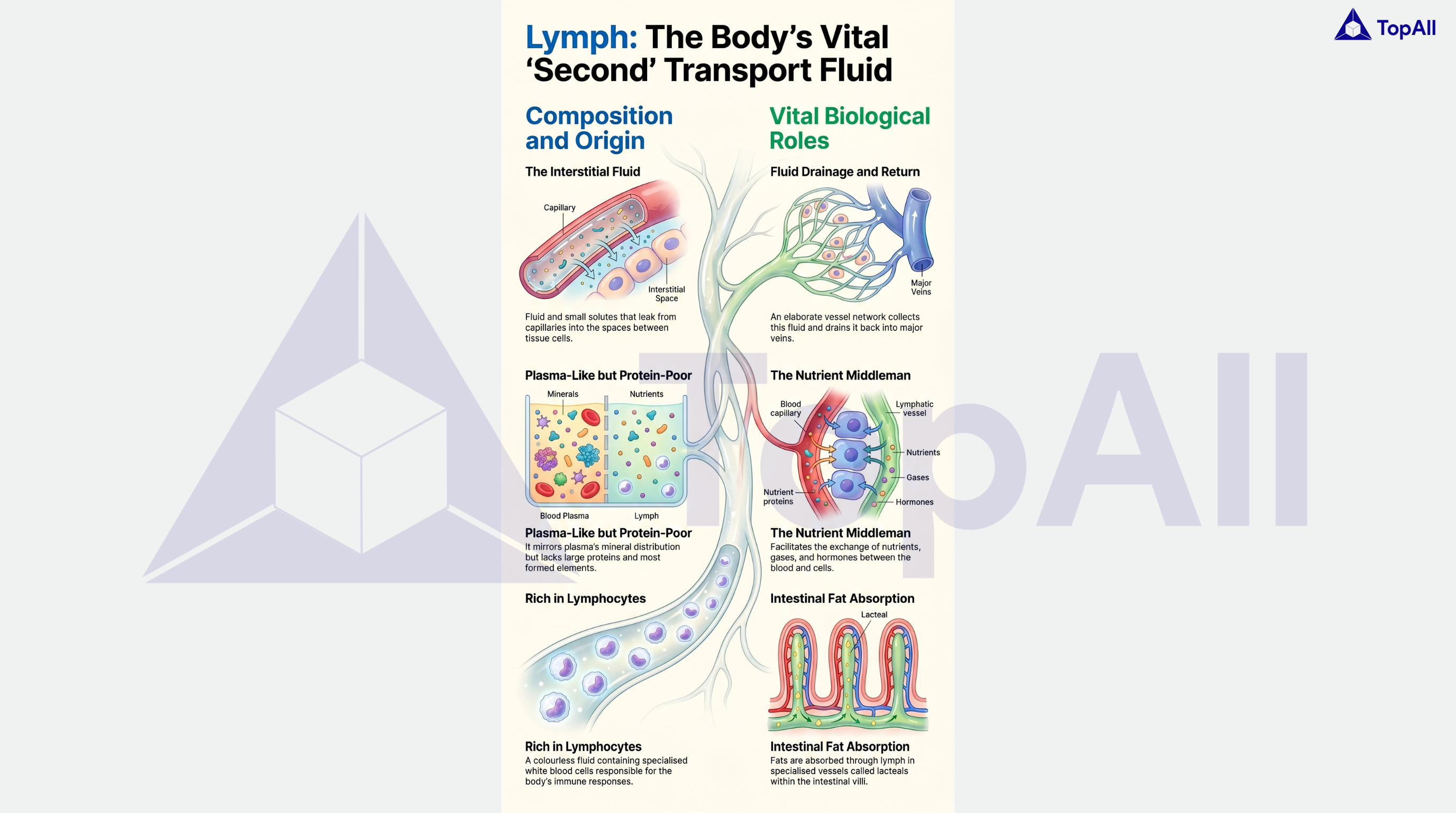
Understand lymph as the body’s second transport fluid for NEET UG with TopAll.
NEET UG · Mind map
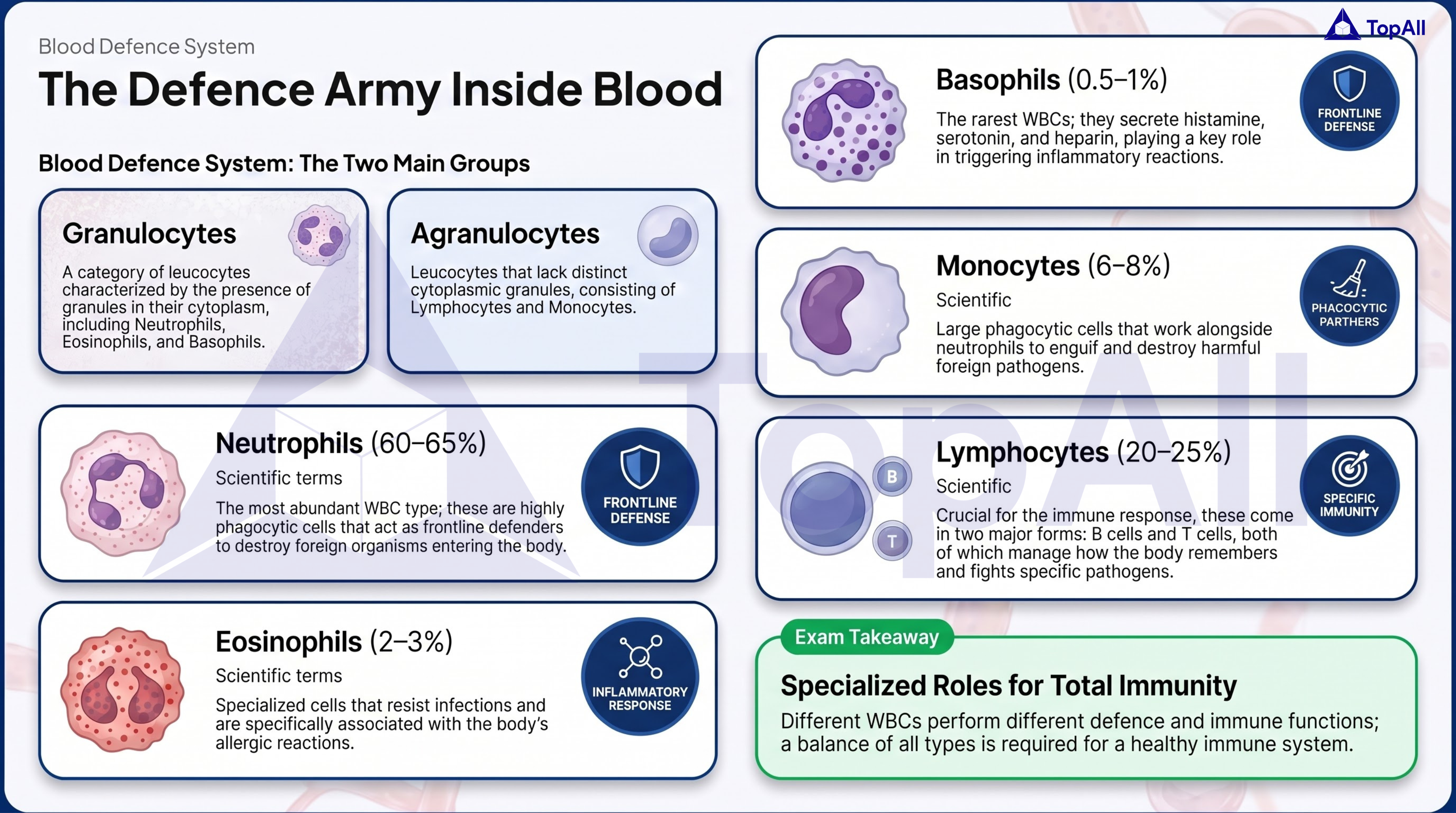
Understand WBC types and immune functions clearly for NEET UG with TopAll.
NEET UG · Mind map
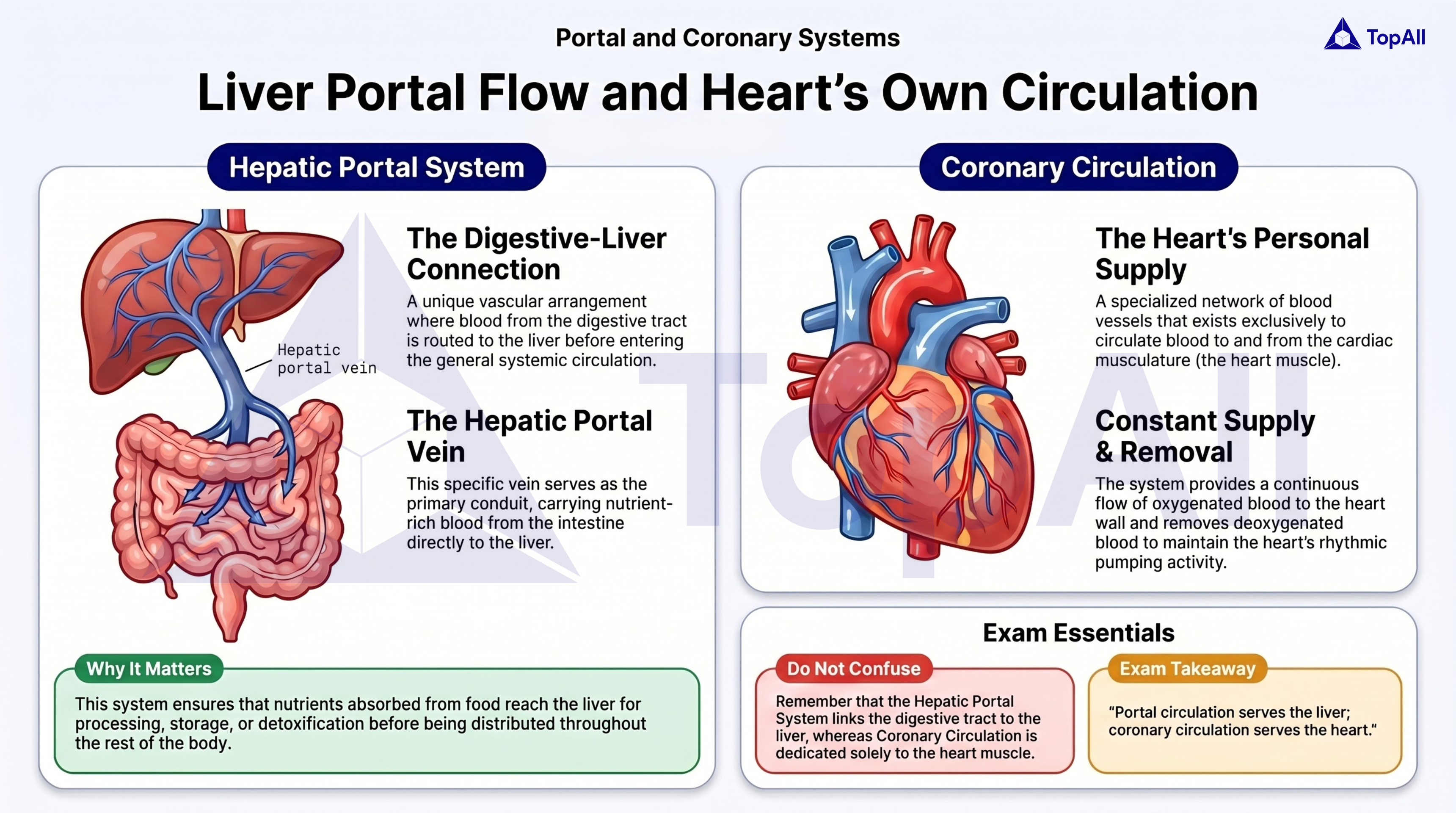
Learn nodal tissue and the heart's pacemaker system for NEET UG with TopAll.
Follow double circulation, distinguish contraction from ejection and calculate cardiac output without confusing vessel names or doubling the flow.
For NEET UG Biology students: trace both circulations without mislabelling pulmonary vessels, use pressure differences to identify valve states and calculate one ventricle’s output without doubling it.
An artery carries blood away from the heart, and a vein carries it toward the heart. Oxygen content is a separate property. In the adult pulmonary circuit, pulmonary arteries carry relatively oxygen-poor blood to the lungs, while pulmonary veins return relatively oxygen-rich blood. The systemic and pulmonary circuits are connected in series through the left and right sides of the heart. A pathway question is best solved by tracing one continuous route.
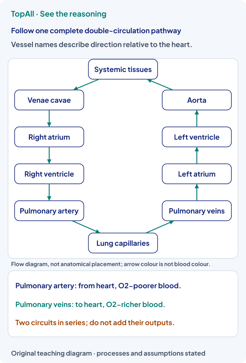
Trace blood leaving a systemic tissue: systemic veins and venae cavae return it to the right atrium; it passes through the tricuspid valve into the right ventricle and then through the pulmonary semilunar valve to the pulmonary artery. After lung exchange, pulmonary veins return it to the left atrium; the mitral valve leads to the left ventricle and the aortic semilunar valve to the aorta. Pulmonary artery and vein names remain correct despite their oxygen-content exception. Valves are crossed only when their phase-specific pressure conditions permit flow.
Atrioventricular valves lie between atria and ventricles: tricuspid on the right, mitral or bicuspid on the left. Semilunar valves guard the pulmonary and aortic outlets. Their opening and closure depend on pressure differences; they do not actively pump blood. Ventricular contraction begins before ejection: during isovolumic contraction, both inlet and outlet valves are closed, so pressure rises without an ejected volume.
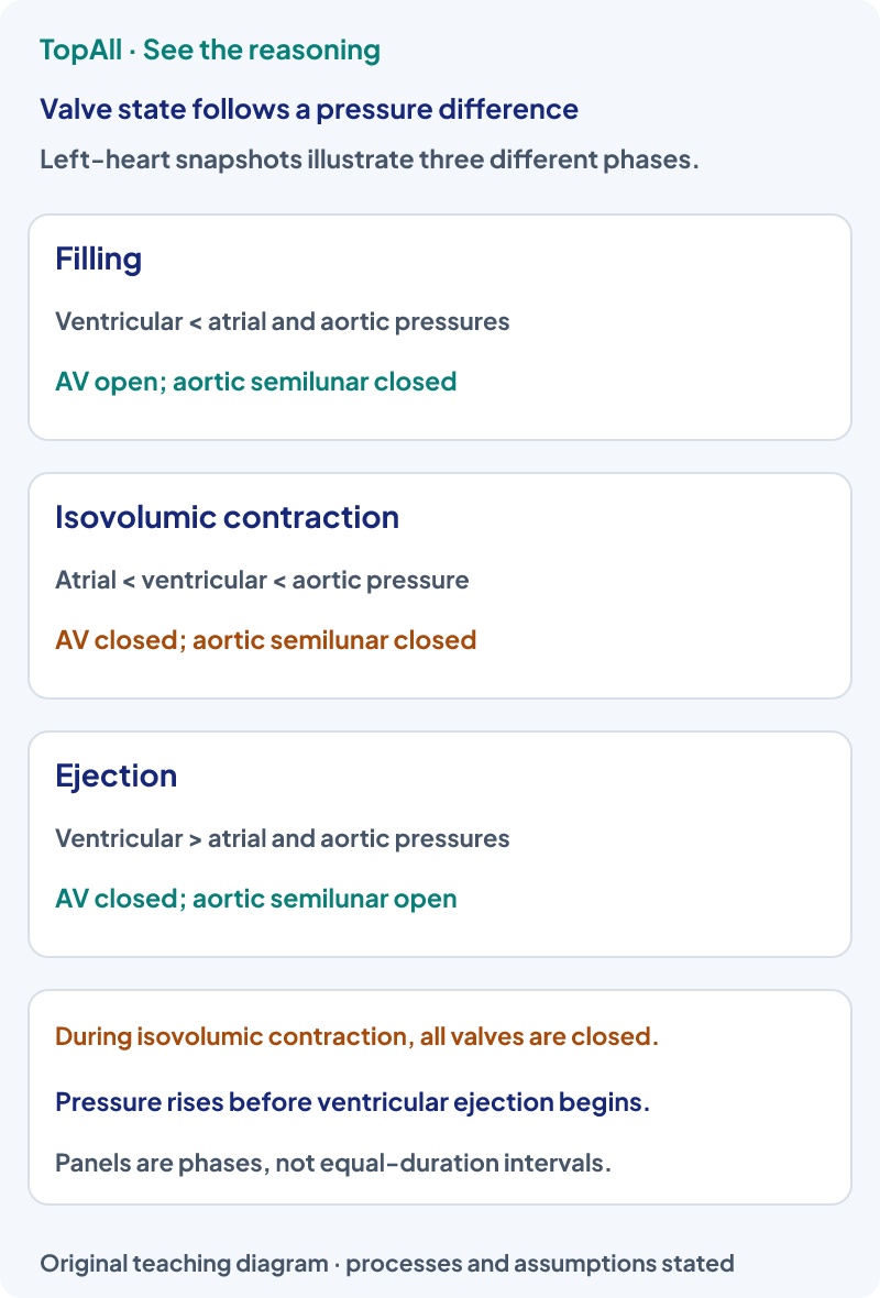
Suppose left atrial pressure is 8, left ventricular pressure 50 and aortic pressure 80 mm Hg in an illustrative early-contraction snapshot. Ventricular pressure exceeds atrial pressure, so the mitral valve is closed. Aortic pressure still exceeds ventricular pressure, so the aortic semilunar valve is also closed. With all valves closed, ventricular volume is temporarily constant while pressure rises. “Ventricle contracting” is therefore insufficient to infer that ejection has already begun.
The sinoatrial node normally initiates the heartbeat. Conduction passes through the atrial system to the AV node and onward through the AV bundle, its branches and Purkinje fibres to ventricular muscle. In an ECG, P represents atrial depolarisation, QRS ventricular depolarisation and T ventricular repolarisation; atrial repolarisation is usually obscured by QRS. Electrical events precede the associated mechanical responses. The first and second heart sounds are associated with AV and semilunar valve closures respectively, not their openings.
With a supplied heart rate of 75 beats/min and stroke volume of 70 mL per beat,
Plasma is the fluid matrix of blood and contains clotting factors. Serum is the fluid remaining after clotting and lacks the clotting factors consumed or removed in that process; it is not simply plasma with all proteins removed. Blood also contains red cells, white cells and platelets. Lymph returns interstitial fluid and participates in transport and immune functions. A diagram of blood flow is not a complete description of exchange with interstitial fluid and lymph.
Trace the route and ask whether a vessel leads away from or toward the heart. Separately classify oxygen content and the relevant exchange bed. For a valve question, compare the pressures on its two sides. For output, use volume per beat times beats per minute and state which ventricle and time scale the calculation describes.
Blood has just left lung capillaries. Which vessels and chambers take it toward the systemic aorta?
It passes through pulmonary veins to the left atrium, through the mitral valve to the left ventricle, then through the aortic semilunar valve to the aorta when the appropriate valves open. Pulmonary veins are veins because they return toward the heart; their relatively high oxygen content does not make them arteries.
Using supplied HR=80 beats/min and SV=65 mL/beat, calculate cardiac output and the average duration of one cycle.
Output is 80×65=5200 mL/min=5.2 L/min per ventricle. An average cycle lasts 60/80=0.75 s. Do not multiply cycle duration by stroke volume to obtain output; the rate is the reciprocal of the cycle duration after units are aligned. These supplied values do not prescribe a universal resting heart rate or stroke volume.
Trace one full circuit and label the four valves in order. At each cardiac phase, write the pressure comparison that opens or closes a valve. Use Breathing and Exchange of Gases to explain the lung exchange step, or Excretory Products and their Elimination to follow renal blood and filtrate separately.
Practice opens the broad Body Fluids and Circulation chapter pool, not a guaranteed cardiac-valve-only set.
Technical review: Codex (automated), 24 October 2026. Definitions and conventions checked against the linked primary textbooks. Numerical examples, self-checks and diagrams are original; calculations and diagram coordinates are checked separately. Human subject review is not claimed. These focused lessons support the existing NEET Biology chapter catalogue and do not claim complete current exam or syllabus coverage.
After a circulation error, check vessel direction, chamber order and valve pressures independently. Confirm the output units before returning to chapter practice.

