NEET UG · Video

Male Reproductive System for NEET Biology
Study the male reproductive system for NEET UG with TopAll.
9 min video

NEET UG · Biology
Revise the concepts. Try a question. Understand your next step.
54 hard questions available · Try answers free. Sign in to save progress or take a quiz.
NEET UG · Biology
Understand the concept, practise questions and check your learning.
Understand
Separate chromosome number from DNA content, distinguish meiotic and differentiation stages and locate fertilisation and implantation. Human reproduction: count the cell stage and locate the event
Read the guidePractise
Try 54 hard chapter MCQs. Untimed; check answers and available explanations as you go.
Practise chapter MCQsCheck your learning
Prepare a timed chapter quiz. Choose the available question count and pace before starting.
Prepare a chapter quizThis is chapter practice, not a full-length NEET mock paper.
These are playable question counts after quality and duplicate checks.
The practice player uses single-correct-answer MCQs. Chapter quizzes are not full-length NEET mock papers. Check the practice guide and official syllabus sources.
Revise before you practise
NEET UG · Video
Study the male reproductive system for NEET UG with TopAll.
9 min video
NEET UG · Video
Understand menstrual hygiene and health basics for NEET UG with TopAll.
4 min video
NEET UG · Video
Learn parturition and lactation in human reproduction for NEET UG with TopAll.
4 min video
NEET UG · PDF notes
Read PDF · 10 pages →Understand menstrual hygiene and health basics for NEET UG with TopAll.
10 pages
NEET UG · PDF notes
Read PDF · 12 pages →Learn parturition and lactation concepts for NEET UG with TopAll.
12 pages
NEET UG · PDF notes
Read PDF · 12 pages →Study pregnancy and embryonic development for NEET UG with TopAll.
12 pages
NEET UG · Mind map
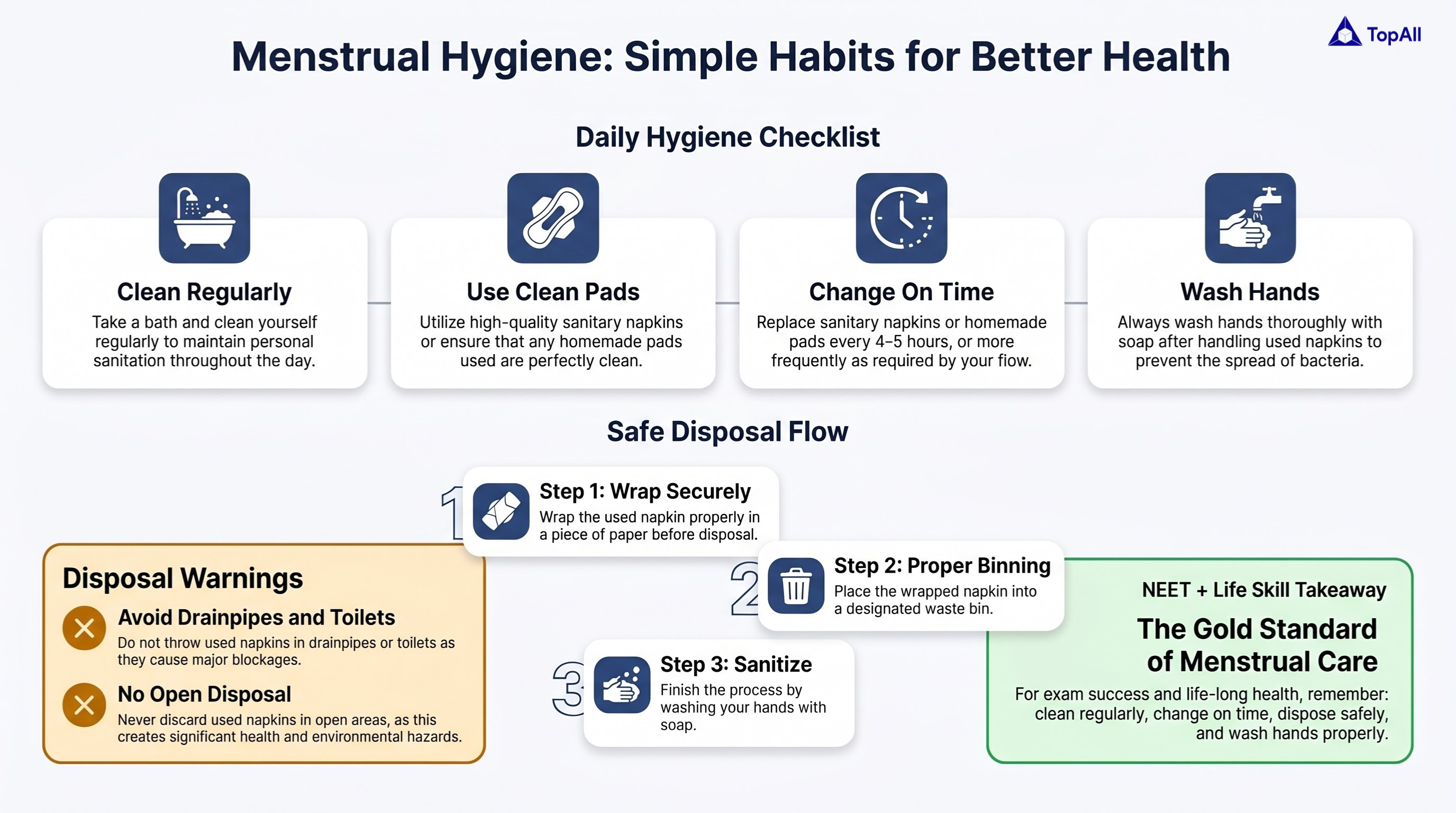
Learn menstrual hygiene and health basics for NEET UG with TopAll.
NEET UG · Mind map
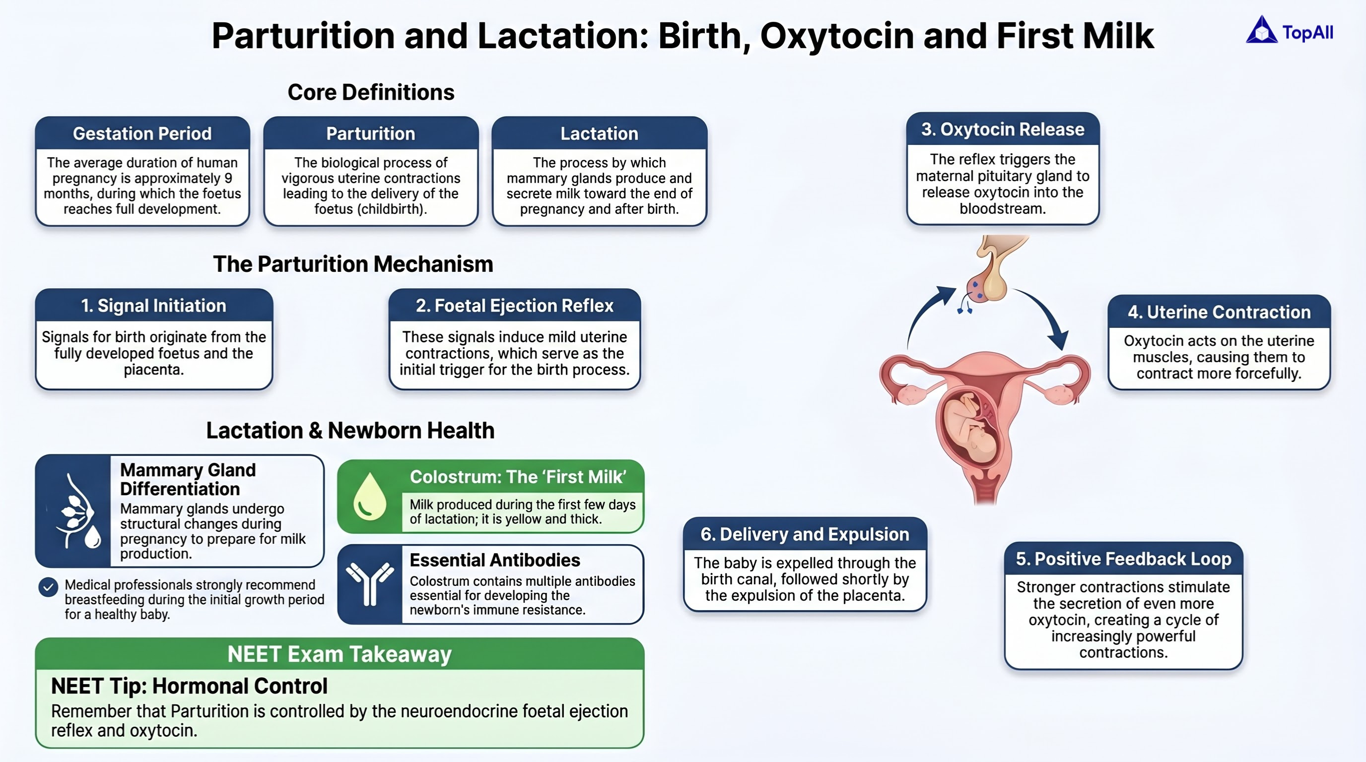
Study parturition and lactation clearly for NEET UG with TopAll.
NEET UG · Mind map
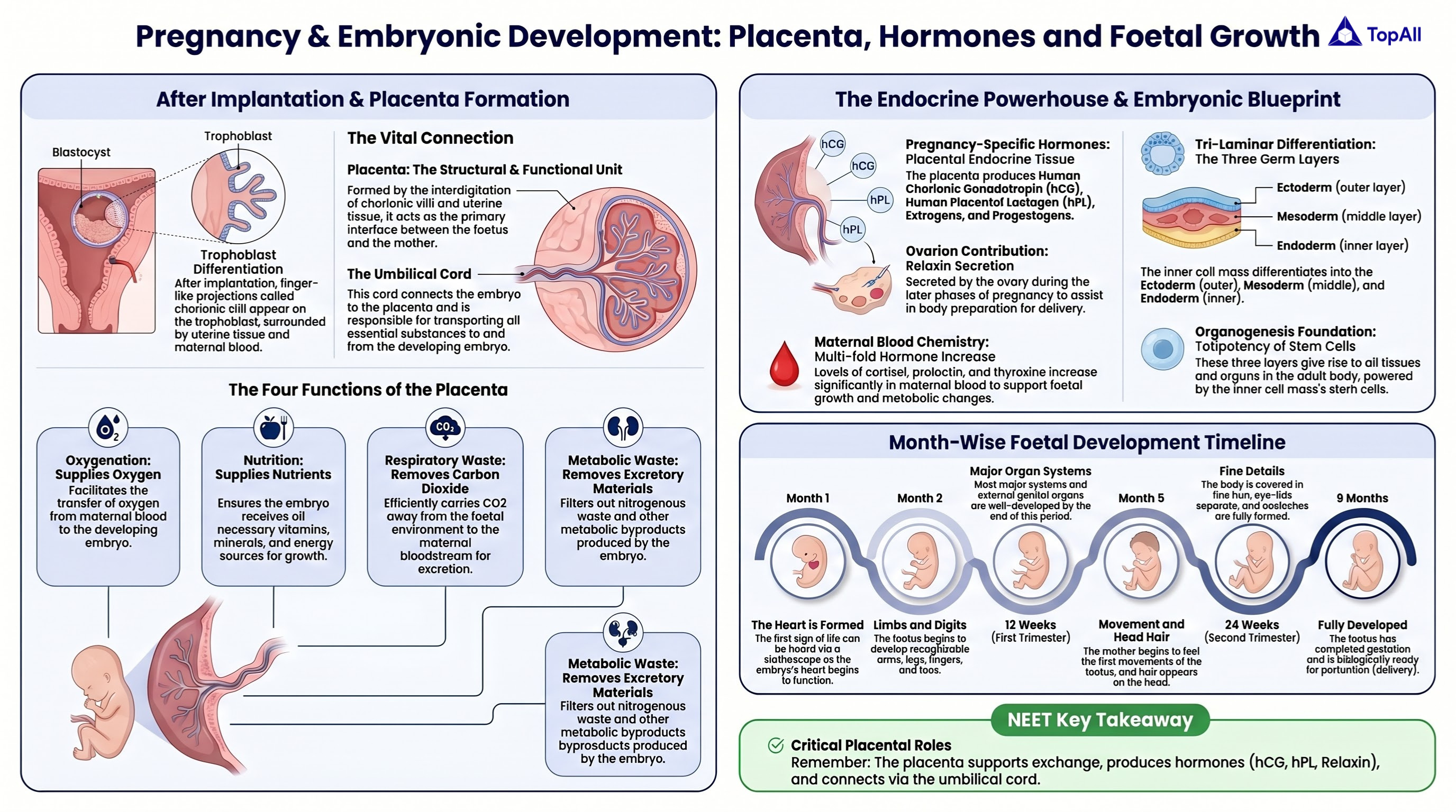
Learn pregnancy and embryonic development for NEET UG with TopAll.
Separate chromosome number from DNA content, distinguish meiotic and differentiation stages and locate fertilisation and implantation.
For NEET UG Biology students: count chromosomes, DNA and cells separately, distinguish meiosis from differentiation and place reproductive events in the correct location.
Count chromosomes by centromeres in the standard model. DNA replication duplicates chromatids without doubling the chromosome number before segregation. A primary spermatocyte after replication is diploid with 46 chromosomes and 4C DNA; a secondary spermatocyte has 23 chromosomes and 2C DNA. A spermatid has 23 chromosomes and 1C DNA. C denotes an unreplicated haploid DNA complement.
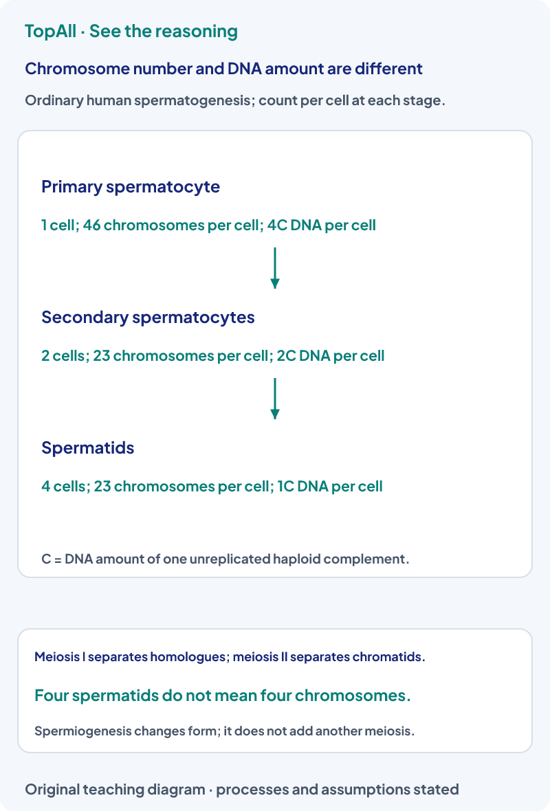
One primary spermatocyte has 4C DNA. It gives two secondary spermatocytes with 2C each, so the two-cell total remains 4C in the supplied complete-division model. Saying each secondary cell has 4C would double-count the initial DNA. Saying the chromosome count stays 46 in each would miss the homologous reduction.
The primary oocyte begins meiosis before birth and remains arrested in prophase I in the standard account. Completion of meiosis I gives a large secondary oocyte and a small polar body. The ovulated secondary oocyte is associated with metaphase-II arrest; fertilisation-associated activation allows completion. Do not apply the four equivalent spermatid output model directly to oogenesis.
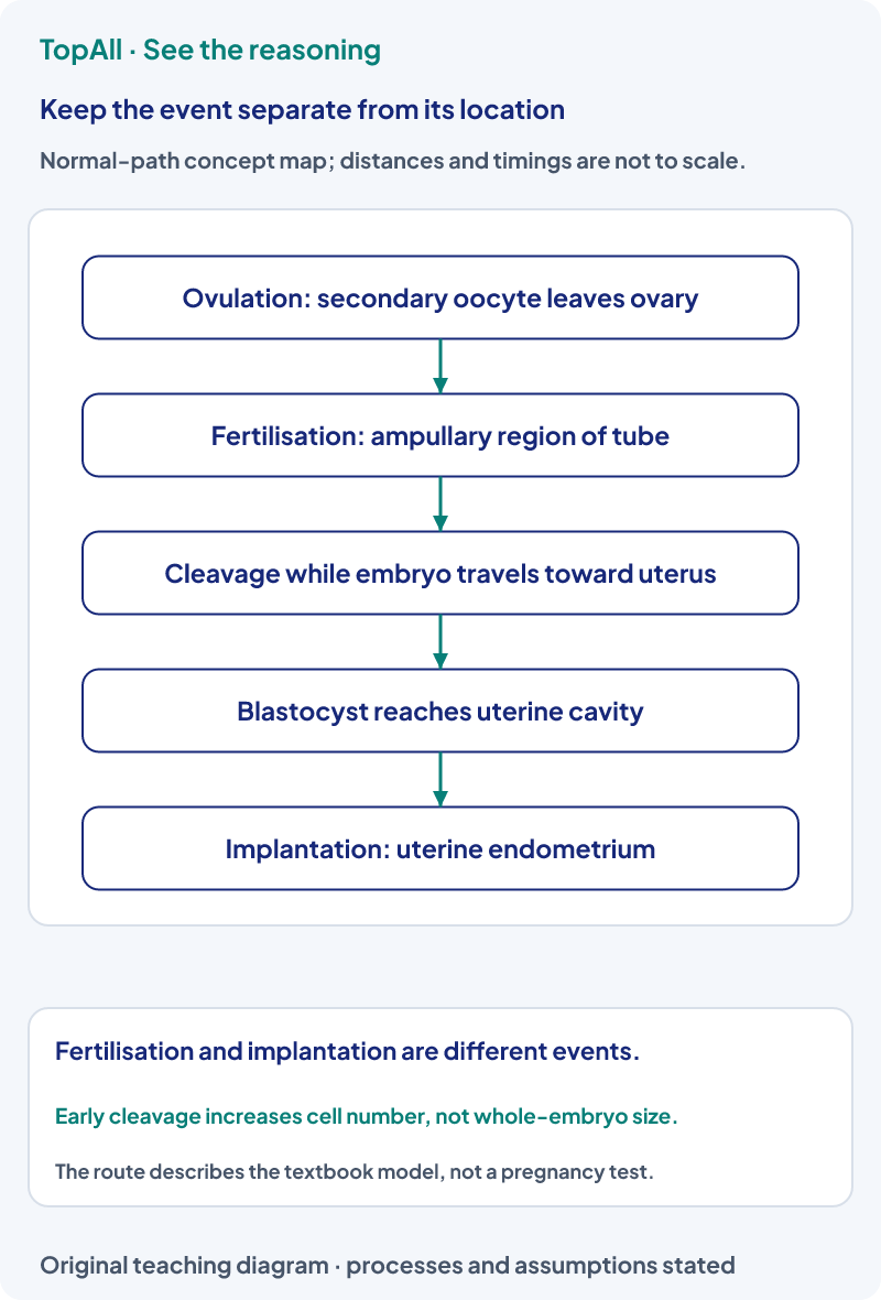
An original explanation says fertilisation normally occurs in the uterine endometrium because that is where the embryo develops later. It has exchanged two stages. Fertilisation belongs to the ampullary region in the cited NCERT normal-path account; implantation involves the uterine endometrium. The later destination does not relocate the earlier event.
Spermatogenesis includes production of sperm through the germ-cell sequence. Spermiogenesis concerns differentiation of spermatids into spermatozoa, and spermiation concerns release from the seminiferous epithelium. Similar word beginnings do not make the processes interchangeable. A shape change is not automatically a reduction in chromosome number.
In an original ideal account, five primary spermatocytes each complete meiosis with all products retained. They yield twenty spermatids, each with23 chromosomes. Twenty is the cell count, not the chromosomes in one cell. The exercise does not predict an individual’s measured sperm count or guarantee survival and maturation of every product.
Early cleavage partitions the initial embryo into more blastomeres while its overall size remains approximately constrained by the surrounding context. Morula and blastocyst name developmental organisation, not just two arbitrary cell totals. The trophoblast and inner cell mass have different roles; naming one does not describe the entire blastocyst.
Write three labels beside an answer: developmental stage, location and what is being counted. A follicle is not simply another word for its oocyte. A secondary oocyte is not diploid merely because each chromosome still has two chromatids. Avoid treating a simplified cycle diagram as a universal personal timetable.
A diploid cell completes DNA replication without chromosome segregation. Has it become tetraploid solely because its DNA doubled?
No. Replication duplicates chromatids while the homologous-set count remains diploid in the stated model. DNA amount and ploidy are different descriptors. The chromosome count stays 46 for the ordinary human diploid cell before sister-centromere separation.
A spermatid changes into a spermatozoon without another meiotic division. Which term names this change, and does it halve 23 chromosomes again?
The change is spermiogenesis. It does not halve the chromosome number to a non-integer value or create another reduction division. The ordinary haploid chromosome count remains 23 during this differentiation account.
Count cells, chromosomes per cell and DNA per cell through meiosis. Then locate fertilisation and implantation separately. Compare assisted-reproduction vocabulary in Reproductive Health, or revisit immune cell versus organ questions in Human Health and Diseases.
Practice opens the broad reproduction chapter pool; this guide is a curriculum model rather than a clinical assessment.
Technical review: Codex (automated), 24 October 2026. Core mechanisms were compared with the linked primary textbooks and university or government references. All diagrams, problem inputs and explanations are original. Numerical balances and diagram directions are checked separately. Independent human subject review is not claimed. These focused guides support the existing chapter catalogue; they do not claim complete current exam or syllabus coverage.
For a counting error, label the denominator per cell or across all products. For a location error, name the event before naming the organ.

