NEET UG · Video

Vertebral Column and Rib Cage
Learn vertebral column and rib cage structure for NEET UG with TopAll.
5 min video
NEET UG · Biology
Revise the concepts. Try a question. Understand your next step.
30 hard questions available · Try answers free. Sign in to save progress or take a quiz.
NEET UG · Biology
Understand the concept, practise questions and check your learning.
Understand
Use matched sarcomere diagrams to explain shortening, preserve filament lengths and place ATP and calcium at the correct steps. Locomotion: read sliding filaments and separate ATP from calcium roles
Read the guidePractise
Try 30 hard chapter MCQs. Untimed; check answers and available explanations as you go.
Practise chapter MCQsCheck your learning
Prepare a timed chapter quiz. Choose the available question count and pace before starting.
Prepare a chapter quizThis is chapter practice, not a full-length NEET mock paper.
These are playable question counts after quality and duplicate checks.
The practice player uses single-correct-answer MCQs. Chapter quizzes are not full-length NEET mock papers. Check the practice guide and official syllabus sources.
Revise before you practise
NEET UG · Video
Learn vertebral column and rib cage structure for NEET UG with TopAll.
5 min video
NEET UG · Video
Compare skeletal, visceral and cardiac muscles for NEET UG with TopAll.
4 min video
NEET UG · Video
Study sliding filament changes during contraction for NEET UG with TopAll.
6 min video
NEET UG · PDF notes
Read PDF · 10 pages →Learn vertebral column and rib cage structure for NEET UG with TopAll.
10 pages
NEET UG · PDF notes
Read PDF · 9 pages →Compare skeletal, visceral and cardiac muscles for NEET UG with TopAll.
9 pages
NEET UG · PDF notes
Read PDF · 10 pages →Study sliding filament changes during contraction for NEET UG with TopAll.
10 pages
NEET UG · Mind map
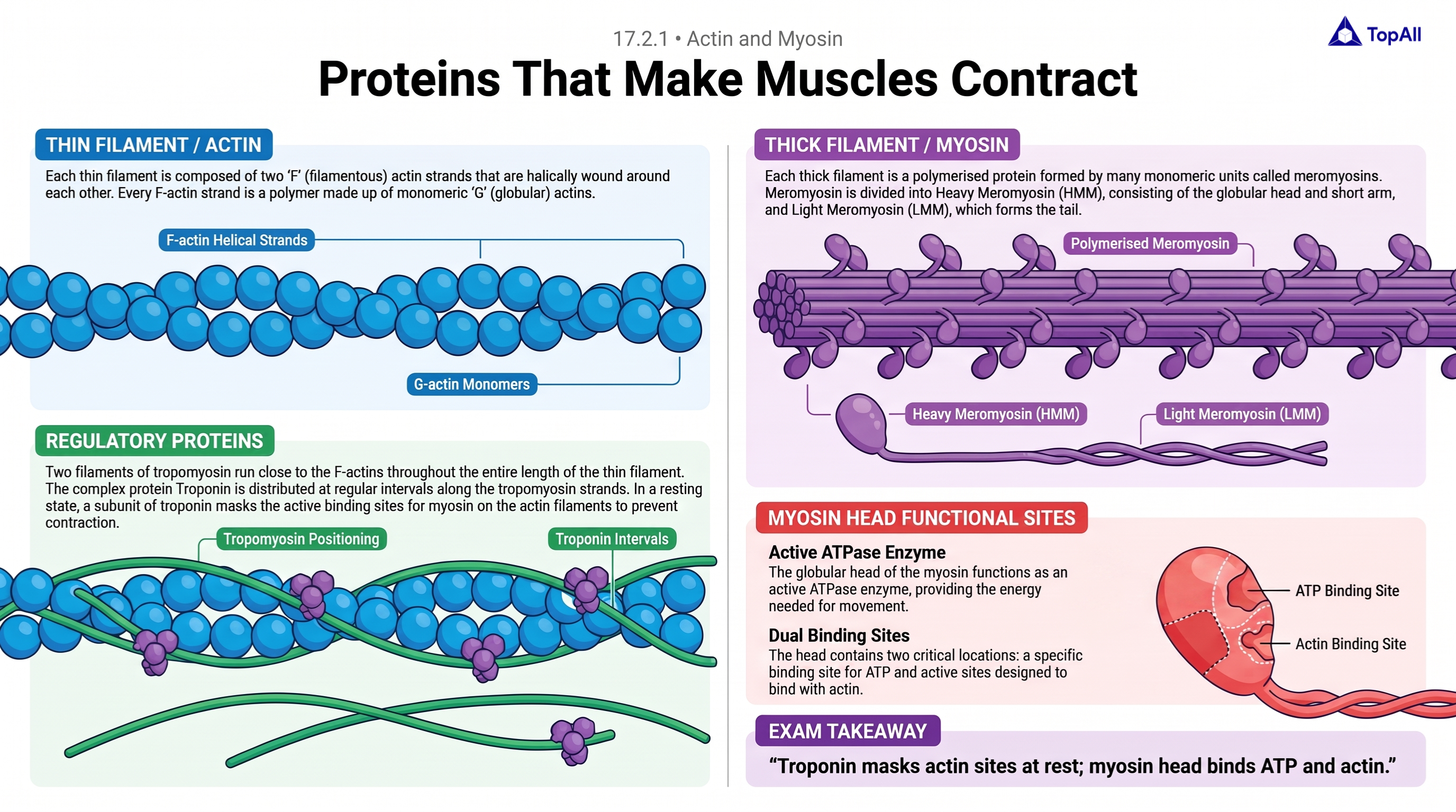
Learn contractile protein structure for NEET UG with TopAll.
NEET UG · Mind map
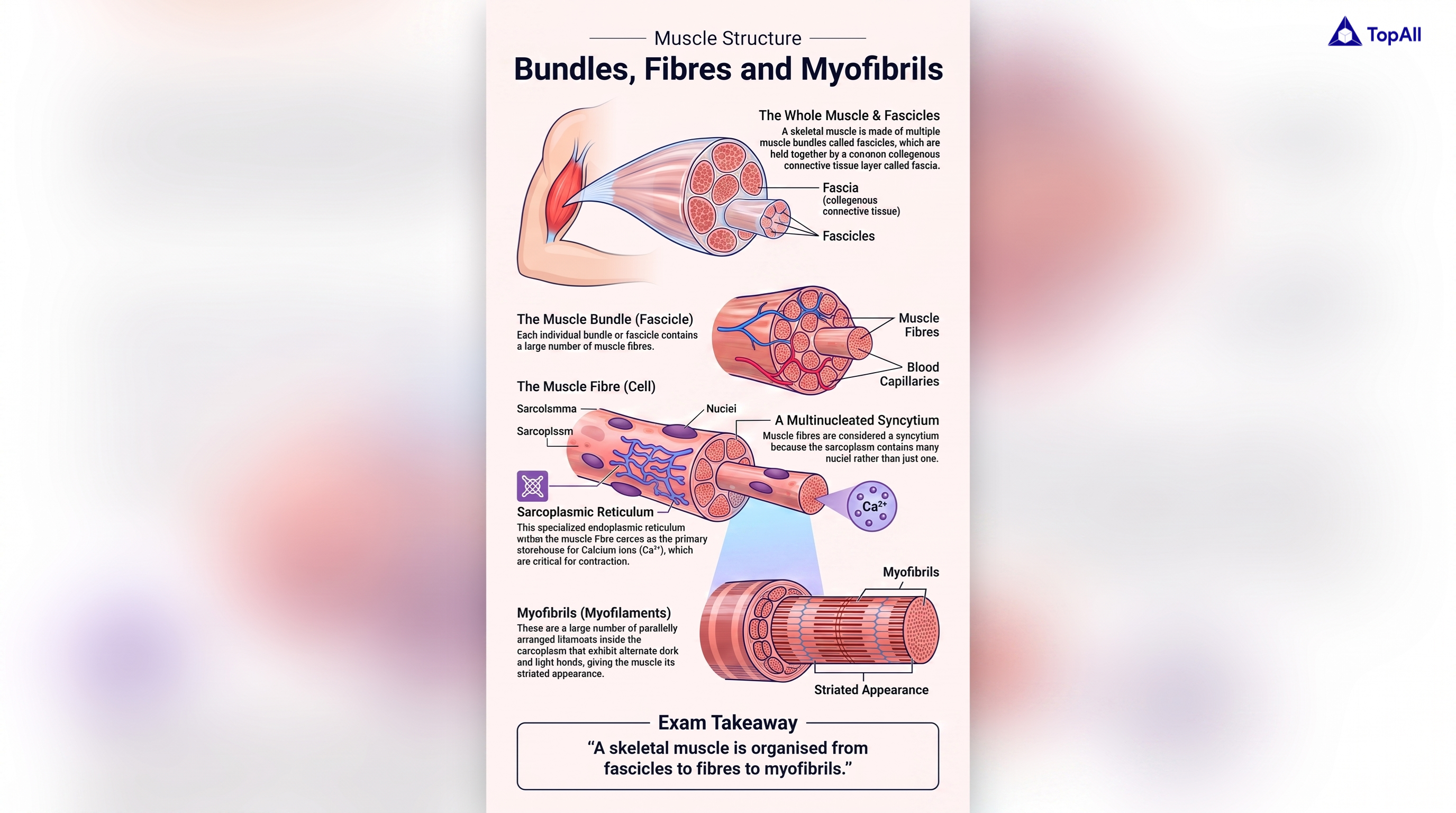
Master skeletal muscle organisation for NEET UG with TopAll.
NEET UG · Mind map
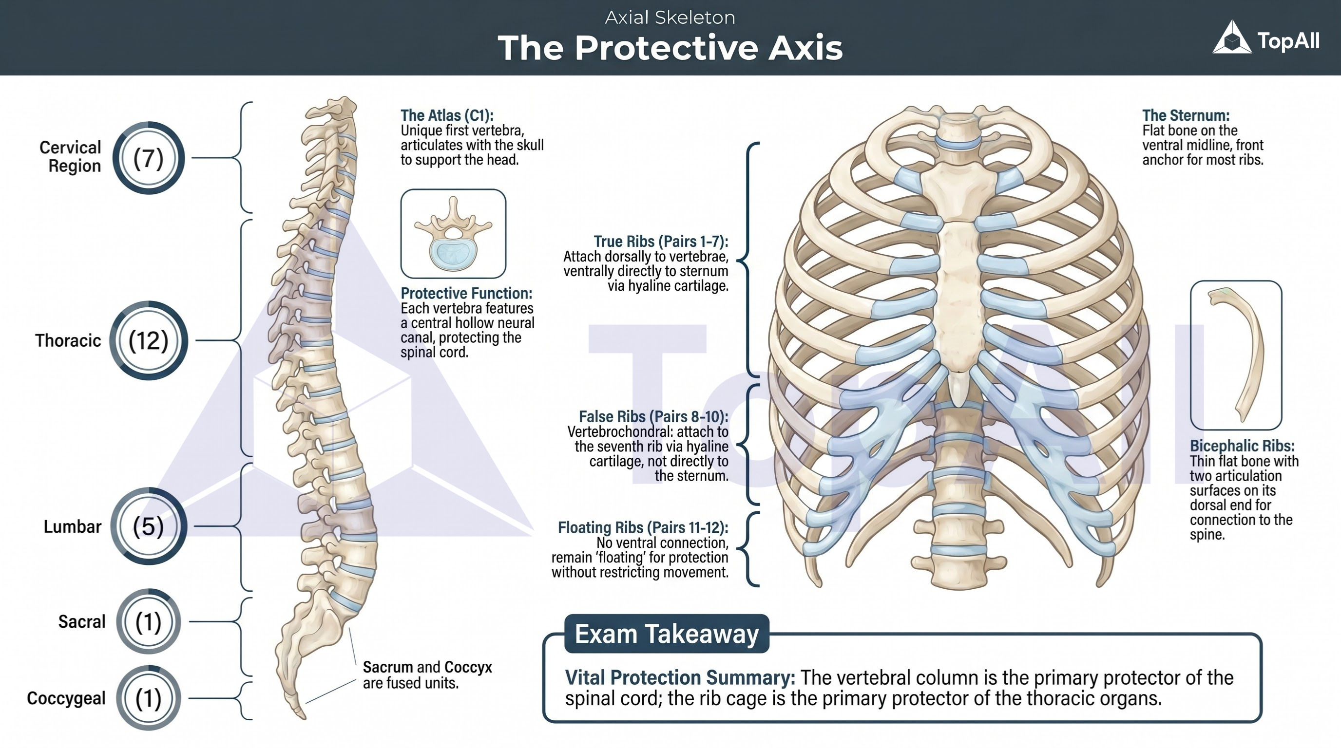
Learn vertebral column and rib cage structure for NEET UG with TopAll.
Use matched sarcomere diagrams to explain shortening, preserve filament lengths and place ATP and calcium at the correct steps.
For NEET UG Biology students: read shortening from sarcomere geometry, identify what stays fixed in the sliding-filament model and distinguish ATP binding from ATP hydrolysis and calcium-dependent access to actin.
A sarcomere extends from one Z line to the next. In the sliding-filament model, thin filaments move relative to thick filaments, bringing Z lines closer. Actin and myosin filaments retain their lengths. The A band corresponds to the thick-filament extent and stays the same length; the I regions and H zone become narrower as overlap increases. A muscle fibre is a cell containing many myofibrils, which contain serial sarcomeres; these are different levels of organisation.
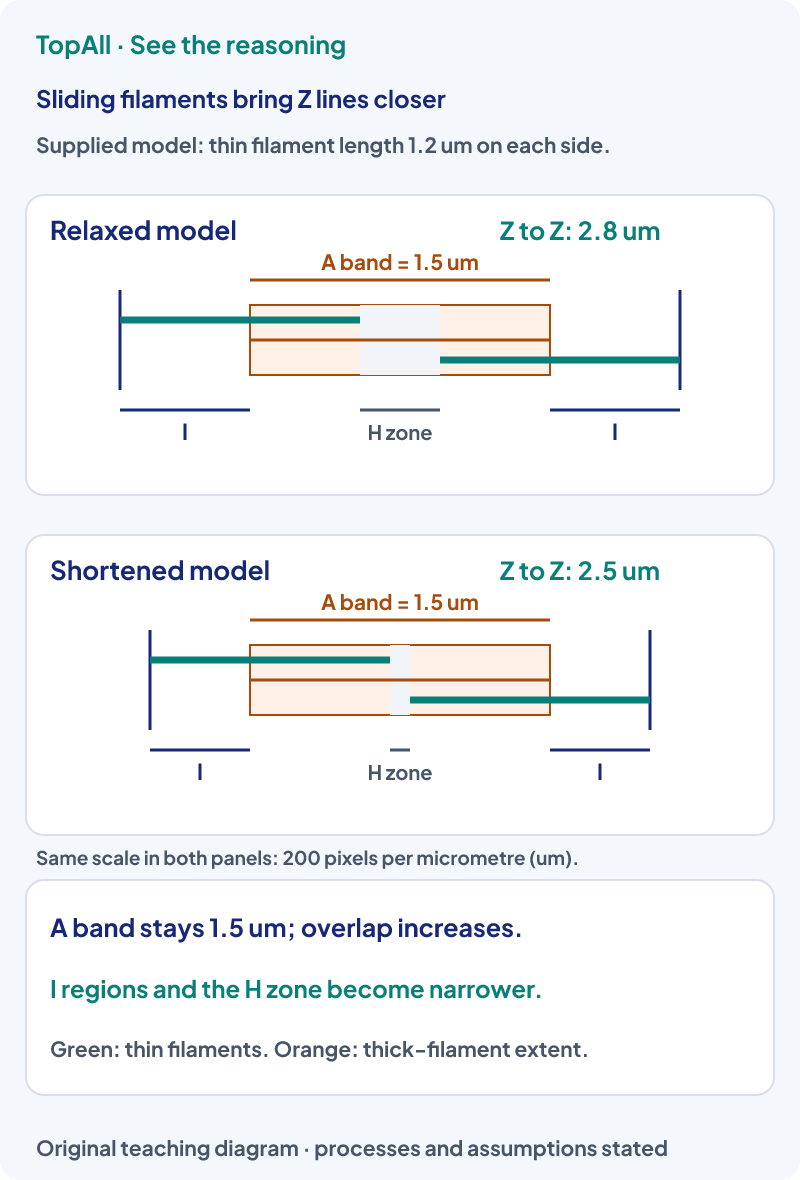
In the supplied model, Z-to-Z length decreases from 2.8 to 2.5 μm: shortening is 0.3 μm, about 10.7% of the starting length. The A band remains 1.5 μm. With a 1.2 μm thin filament anchored at each end and no central thin-filament overlap, H-zone width is L−2(1.2): initially 0.4 μm and finally 0.1 μm. The total thin-only I extent is L−1.5: 1.3 then 1.0 μm. These geometric results concern the specified model, not universal dimensions of all sarcomeres.
At the skeletal neuromuscular junction, a motor signal and acetylcholine trigger muscle-membrane excitation and release of from the sarcoplasmic reticulum into sarcoplasm. Calcium binds troponin, allowing tropomyosin to move so myosin-binding sites on actin become accessible. ATP supports cross-bridge cycling and calcium recovery. Do not substitute “calcium binds myosin to supply energy” for the distinct roles of troponin, actin access and ATP.
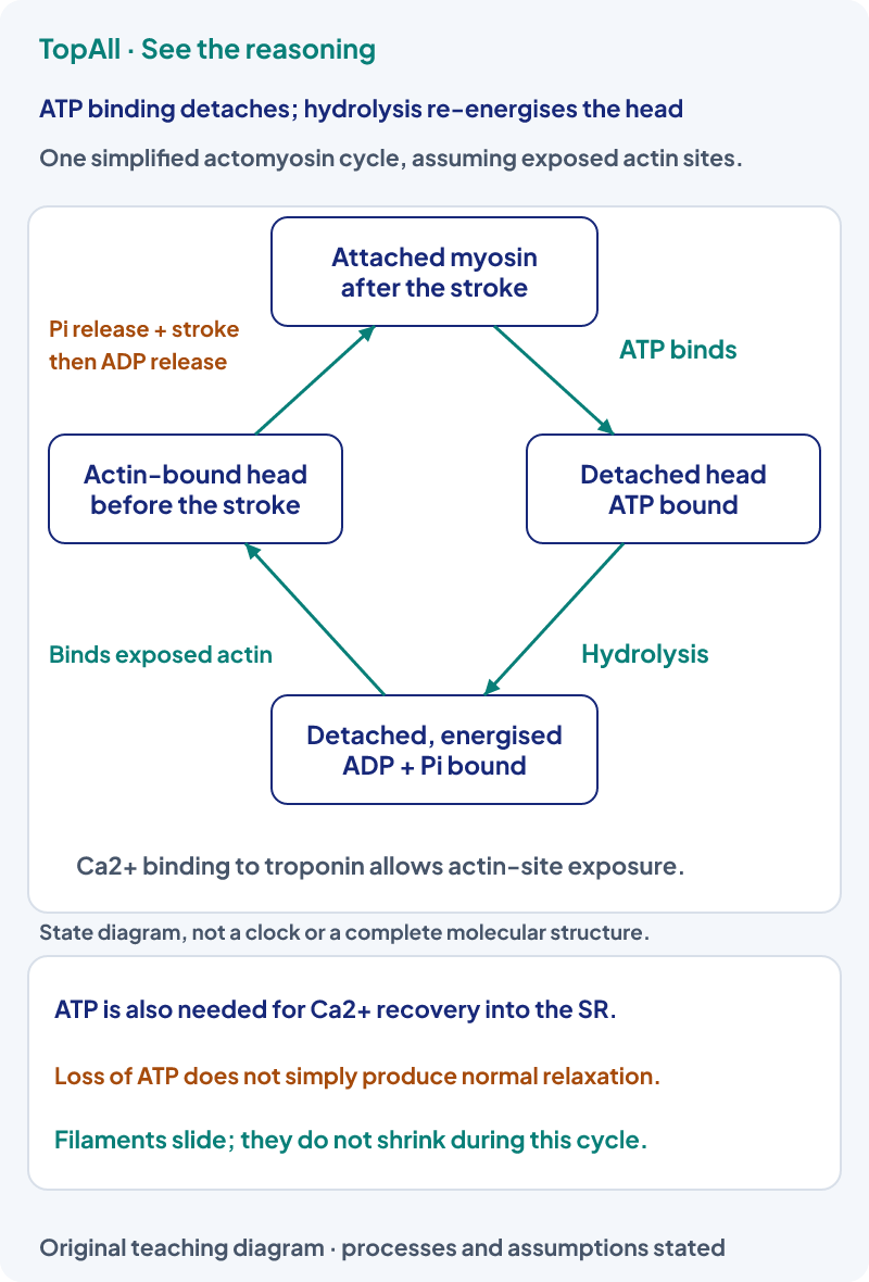
A myosin head remains attached after a stroke. A new ATP molecule binds, allowing that cross-bridge to detach. Hydrolysis then re-energises the detached head for another cycle, provided actin sites can be accessed. Thus an option saying “ATP binding causes detachment” describes a different step from “ATP hydrolysis re-energises the head”. Without ATP, a head cannot simply execute normal detachment and relaxation; ATP also supports pumping calcium back into the SR.
A muscle develops tension by contraction; it does not actively push a bone in the opposite direction. Flexor and extensor groups can act antagonistically, with coordinated activation producing the desired joint movement. In a withdrawal response, inhibitory spinal pathways can reduce activation of antagonist muscles. “Voluntary skeletal muscle” describes its control category; it does not mean that every skeletal-muscle action requires a conscious decision.
Suppose ten identical sarcomeres arranged end to end each change from 2.8 to 2.5 μm, with no extra end gaps in this simplified chain. Total length changes from 28 to 25 μm, so the chain shortens by 3 μm, still about 10.7%. Adding more units in series adds their length changes; it does not multiply the percentage shortening by ten. Real muscle geometry includes many parallel myofibrils, connective tissues and attachments beyond this simplified length model.
Tendons connect skeletal muscle to bone, while ligaments connect bone to bone across joints. A joint’s structure limits its possible motion: the elbow is a standard hinge example, the shoulder a ball-and-socket example and the atlas-axis articulation a pivot example. These examples identify structural patterns; they do not make every named movement exclusive to one isolated muscle or one tissue layer.
Locate both Z lines and thick-filament ends before comparing panels. Keep filament length fixed and calculate overlap from the stated geometry. Separate calcium-dependent exposure of binding sites from ATP-dependent cycling. In a chain, distinguish an absolute length change from a percentage. Name tendon, ligament and joint type from their actual connections.
In a standard sliding-filament comparison, sarcomere length decreases while thin and thick filament lengths are fixed. Which changes: A band, I regions, H zone or all three in the same way?
The A-band length stays fixed because it represents thick-filament length. The I regions and H zone narrow as overlap increases; the H zone can disappear if overlap becomes sufficient. Thin and thick filaments do not shrink. The exact numerical changes require the starting geometry rather than an assumption that every band shortens equally.
A fibrous structure joins muscle to bone, and another joins one bone to another across a joint. Name each and its broad tissue category.
Muscle-to-bone is a tendon; bone-to-bone is a ligament. Both are specialised connective tissues with extracellular fibres, rather than epithelial sheets or contractile muscle cells. Their connections help identify their roles. This structural classification is separate from whether a nearby joint is hinge, pivot or ball-and-socket.
Mark Z lines, thick-filament ends and thin-filament ends. State what moves and what keeps its length, then identify the ATP and calcium steps. Use Neural Control and Coordination to trace the initiating signal, or Structural Organisation in Animals to distinguish muscle, tendon and the other tissues in an organ.
Practice opens the broad Locomotion and Movement chapter pool, not a guaranteed sarcomere-only set.
Technical review: Codex (automated), 24 October 2026. Definitions and conventions checked against the linked primary textbooks. Numerical examples, self-checks and diagrams are original; calculations and diagram coordinates are checked separately. Human subject review is not claimed. These focused lessons support the existing NEET Biology chapter catalogue and do not claim complete current exam or syllabus coverage.
After a movement error, check organisation level, fixed filament lengths, ATP step and attachment type before returning to chapter practice.

