NEET UG · Video

Sexual Dimorphism in Frogs NEET Guide
Understand sexual dimorphism in frogs with examples for NEET UG with TopAll.
4 min video

NEET UG · Biology
Revise the concepts. Try a question. Understand your next step.
78 hard questions available · Try answers free. Sign in to save progress or take a quiz.
NEET UG · Biology
Understand the concept, practise questions and check your learning.
Understand
Classify epithelia from layers and shape, separate tissues from organs and compare the NCERT adult frog’s forelimb and hindlimb digit patterns. Animal structure: read tissue evidence and keep the organism-specific pattern
Read the guidePractise
Try 78 hard chapter MCQs. Untimed; check answers and available explanations as you go.
Practise chapter MCQsCheck your learning
Prepare a timed chapter quiz. Choose the available question count and pace before starting.
Prepare a chapter quizThis is chapter practice, not a full-length NEET mock paper.
These are playable question counts after quality and duplicate checks.
The practice player uses single-correct-answer MCQs. Chapter quizzes are not full-length NEET mock papers. Check the practice guide and official syllabus sources.
Revise before you practise
NEET UG · Video
Understand sexual dimorphism in frogs with examples for NEET UG with TopAll.
4 min video
NEET UG · Video
Learn male frog reproductive system clearly for NEET UG with TopAll.
4 min video
NEET UG · Video
Master frog respiratory system and breathing for NEET UG with TopAll.
4 min video
NEET UG · PDF notes
Read PDF · 7 pages →Understand sexual dimorphism in frogs with examples for NEET UG with TopAll.
7 pages
NEET UG · PDF notes
Read PDF · 9 pages →Learn male frog reproductive system clearly for NEET UG with TopAll.
9 pages
NEET UG · PDF notes
Read PDF · 8 pages →Master frog respiratory system and breathing for NEET UG with TopAll.
8 pages
NEET UG · Mind map
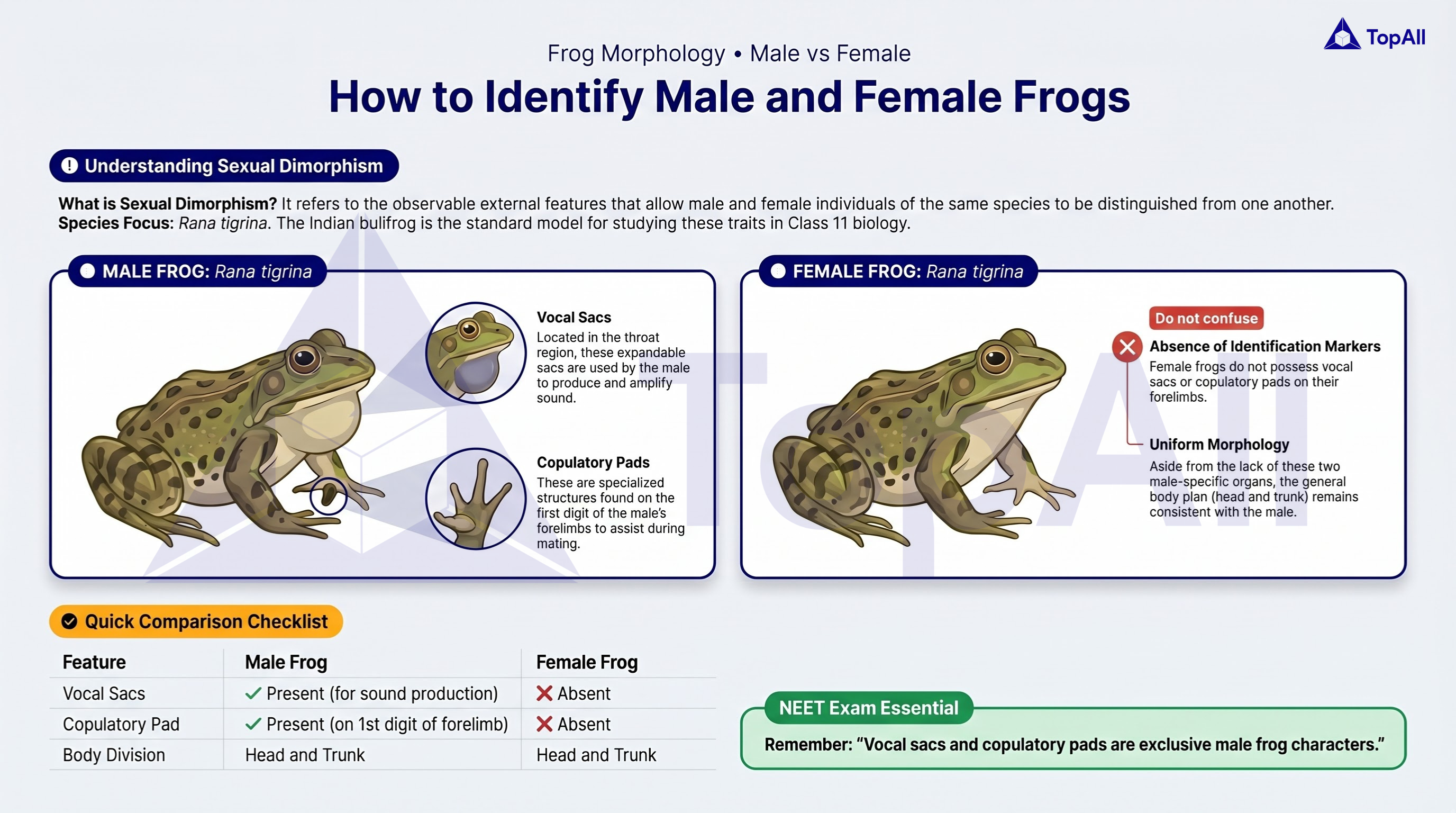
Understand sexual dimorphism in frogs with examples for NEET UG with TopAll.
NEET UG · Mind map
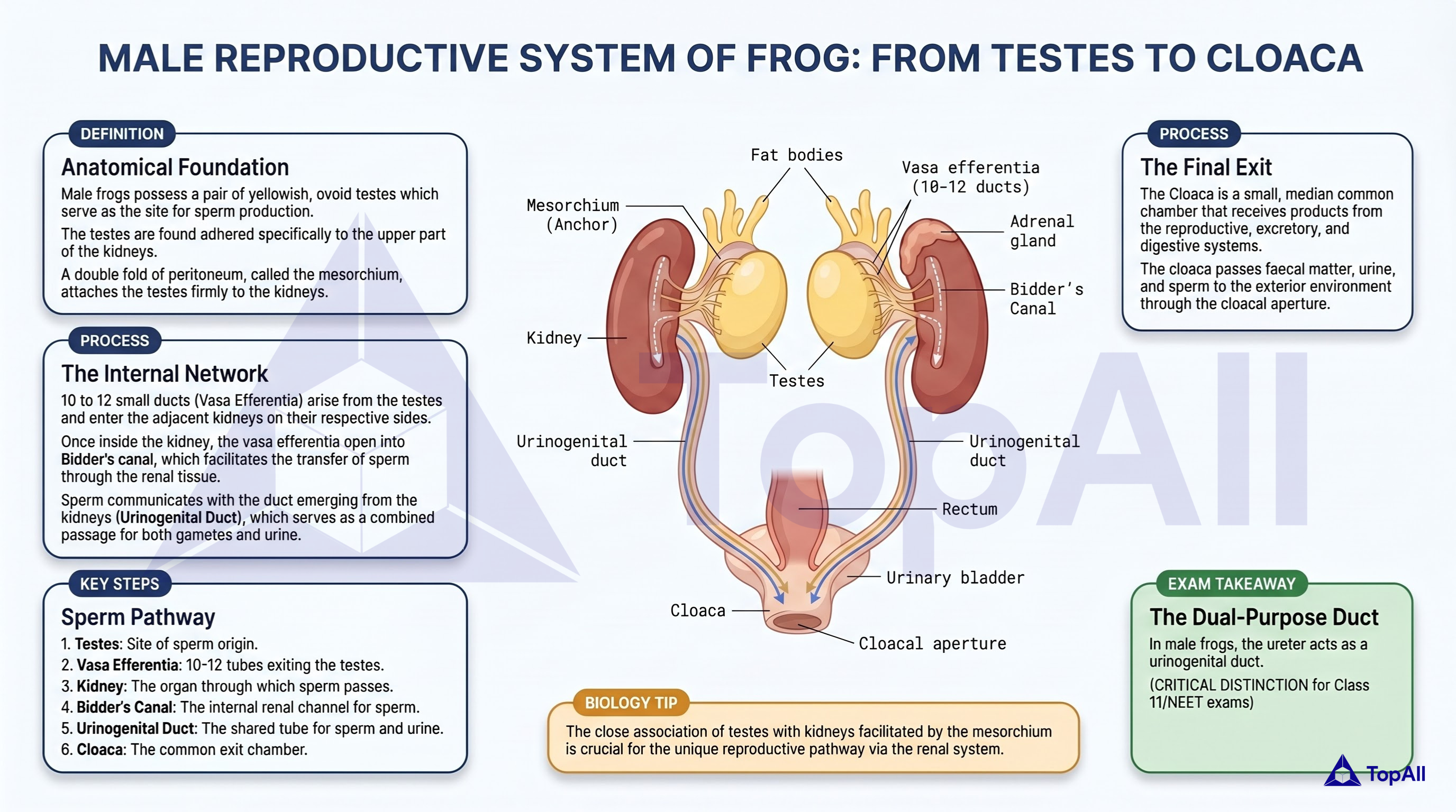
Learn male frog reproductive system clearly for NEET UG with TopAll.
NEET UG · Mind map
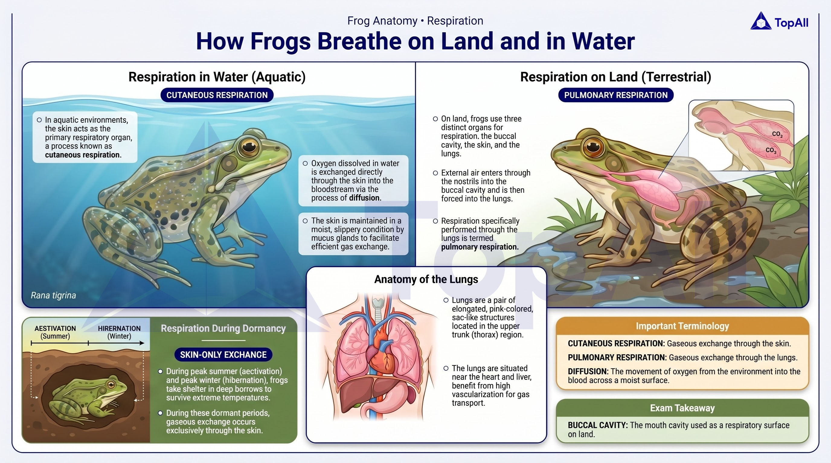
Master frog respiratory system and breathing for NEET UG with TopAll.
Classify epithelia from layers and shape, separate tissues from organs and compare the NCERT adult frog’s forelimb and hindlimb digit patterns.
For NEET UG Biology students: classify a tissue from layer arrangement and cell shape, separate an organ from its lining tissue and use organism-specific external features in the NCERT adult frog example.
Cells and intercellular material form tissues; different tissues combine in organs, and organs interact in systems. The four broad animal tissue classes are epithelial, connective, muscular and neural. In an epithelial image, first establish the number of layers and then the relevant cell shape. A tissue’s likely function helps interpretation, but one function is not a unique identifier. A property of an organ’s lining is not automatically a property of its entire wall.
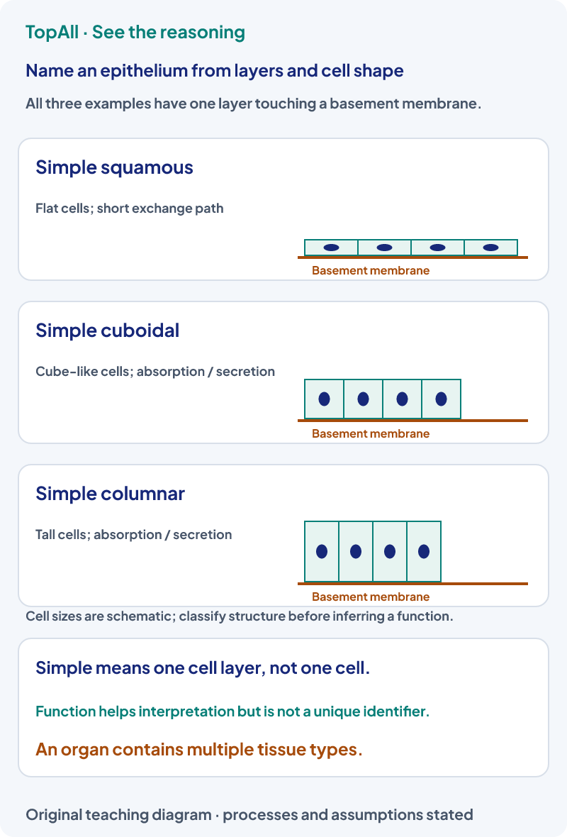
A capillary lining is described as one layer of flattened cells on a basement membrane. The layer-and-shape evidence identifies simple squamous epithelium; in vessels it is called endothelium. The blood within the lumen is a fluid connective tissue, not an epithelial sheet. The short exchange path is consistent with the lining’s role but is supporting information, not a substitute for the structural observations.
For a true stratified epithelium, naming commonly uses the shape of the superficial or apical cells rather than only the basal ones. Pseudostratified epithelium can show nuclei at different heights even though every cell contacts the basal lamina; some cells do not reach the free surface. Thus counting rows of nuclei alone can be misleading. Inspect boundaries, basal contact and apical organisation instead of guessing layer number from one visual cue.
A prompt describes the absorptive lining of small intestine and then asks whether its wall contains only columnar epithelial cells. The absorptive lining is largely simple columnar epithelium with specialised cells, but the whole organ also includes connective tissue, muscle, nerves and vessels. “Simple columnar” answers the specified lining question, not the composition of every part of the intestinal wall. A villus likewise contains more than its surface epithelium.
Connective tissues differ in cells and extracellular components. Blood has a fluid matrix, cartilage has a supportive matrix with embedded cells, and dense fibrous tissues provide tensile connections. Tendons connect muscle to bone and ligaments connect bone to bone. These are connective-tissue examples even though they differ markedly in appearance. An abundance of matrix is a different structural cue from the closely packed cells in a surface epithelium.
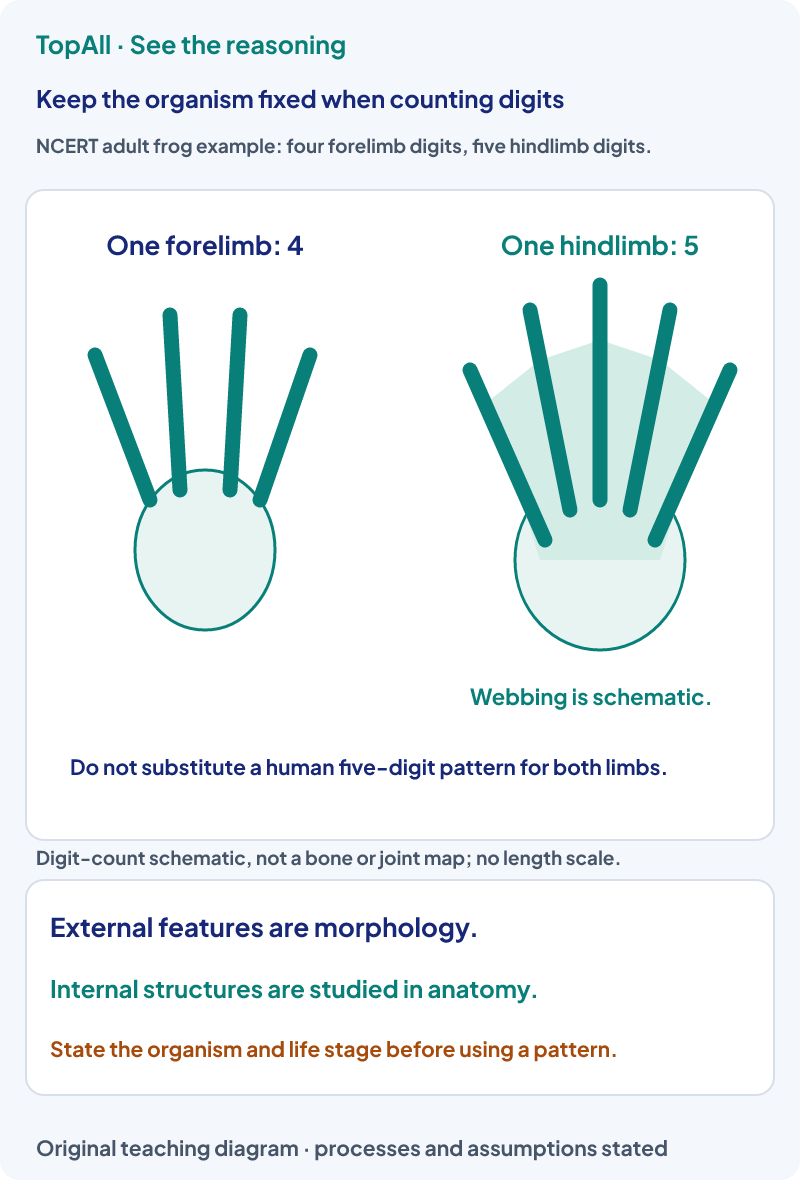
An adult specimen in the textbook frog example has four forelimb digits and five webbed hindlimb digits. A student writes five on both because that is familiar from a human hand and foot. The correction is to keep the organism fixed and use the frog’s observed pattern. The external tympanum is another frog surface feature; it is not a mammalian external ear pinna. The example is a morphological comparison, not a complete identification key for every amphibian species.
Morphology describes external form in this animal comparison, while anatomy examines internal structures. In the NCERT frog example, the heart has two atria and one ventricle, unlike a mammalian four-chambered heart. Frog respiratory surfaces include moist skin and, on land, buccal lining and lungs. The animal’s habitat and life stage matter. These internal and functional observations should not be inferred solely from how many digit rays appear in a limb sketch.
State whether the question names a cell, tissue, organ or organism. For an epithelium, inspect basal contact and surface cell shape. For a connective tissue, inspect matrix and connections. For animal morphology, retain the named organism and life stage; do not replace its features with a familiar mammalian pattern.
An epithelium has nuclei at different heights, but every cell contacts the basal lamina and some do not reach the free surface. Must it be truly stratified?
No. Those observations fit pseudostratified epithelium: the nuclei create an appearance of layers while basal contact shows one layer. A truly stratified arrangement has cells stacked so not all contact the basal lamina. The naming decision should use structural boundaries and contacts, not nuclei height alone.
One structure joins skeletal muscle to bone and another joins bone to bone. Are these whole organ systems, epithelial sheets or specialised connective tissues?
They are specialised connective tissues: tendon for muscle-to-bone and ligament for bone-to-bone. Their fibrous extracellular matrix supports tensile connections. These tissue names do not by themselves name an entire locomotor system, a joint type or the shape of a lining epithelium.
For a tissue, name the layer arrangement and cell or matrix feature that supports your answer. For a frog structure, state the organism and whether the observation is external or internal. Use Digestion and Absorption to connect the intestinal lining to transport, or Locomotion and Movement to connect tendon, ligament and muscle to movement.
Practice opens the broad Structural Organisation in Animals chapter pool, not a guaranteed frog-digit-only set.
Technical review: Codex (automated), 24 October 2026. Definitions and conventions checked against the linked primary textbooks. Numerical examples, self-checks and diagrams are original; calculations and diagram coordinates are checked separately. Human subject review is not claimed. These focused lessons support the existing NEET Biology chapter catalogue and do not claim complete current exam or syllabus coverage.
After a structural-identification error, check organisation level, layer evidence, matrix and the named organism before returning to chapter practice.

