NEET UG · Video

Myelinated and Non Myelinated Fibres
Compare myelinated and non-myelinated nerve fibres for NEET UG with TopAll.
4 min video

NEET UG · Biology
Revise the concepts. Try a question. Understand your next step.
74 hard questions available · Try answers free. Sign in to save progress or take a quiz.
NEET UG · Biology
Understand the concept, practise questions and check your learning.
Understand
Follow sensory and motor pathways, distinguish ion-channel flow from pump maintenance and reason through a chemical-synapse release step. Neural control: trace the pathway and separate ion channels from the pump
Read the guidePractise
Try 74 hard chapter MCQs. Untimed; check answers and available explanations as you go.
Practise chapter MCQsCheck your learning
Prepare a timed chapter quiz. Choose the available question count and pace before starting.
Prepare a chapter quizThis is chapter practice, not a full-length NEET mock paper.
These are playable question counts after quality and duplicate checks.
The practice player uses single-correct-answer MCQs. Chapter quizzes are not full-length NEET mock papers. Check the practice guide and official syllabus sources.
Revise before you practise
NEET UG · Video
Compare myelinated and non-myelinated nerve fibres for NEET UG with TopAll.
4 min video
NEET UG · Video
Learn hindbrain parts and functions for NEET UG with TopAll.
4 min video
NEET UG · Video
Understand midbrain structure and role in coordination for NEET UG with TopAll.
3 min video
NEET UG · PDF notes
Read PDF · 9 pages →Compare myelinated and non-myelinated nerve fibres for NEET UG with TopAll.
9 pages
NEET UG · PDF notes
Read PDF · 9 pages →Learn hindbrain parts and functions for NEET UG with TopAll.
9 pages
NEET UG · PDF notes
Read PDF · 8 pages →Understand midbrain structure and role in coordination for NEET UG with TopAll.
8 pages
NEET UG · Mind map
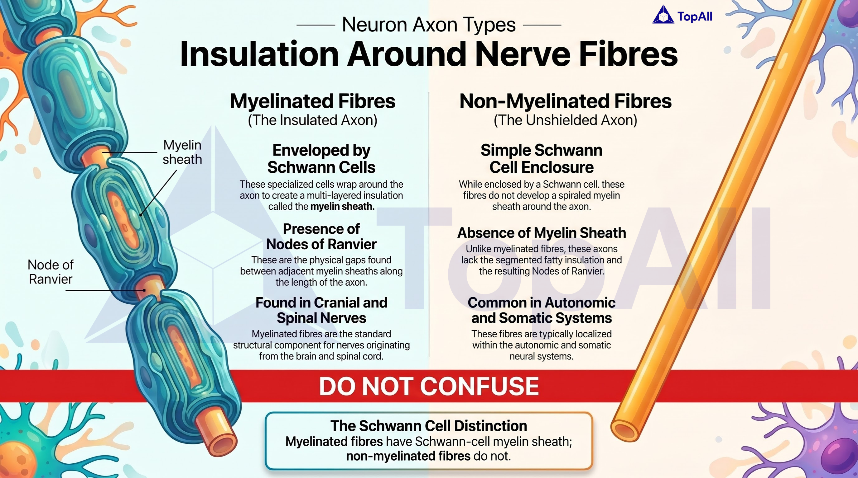
Compare myelinated and non-myelinated nerve fibres for NEET UG with TopAll.
NEET UG · Mind map
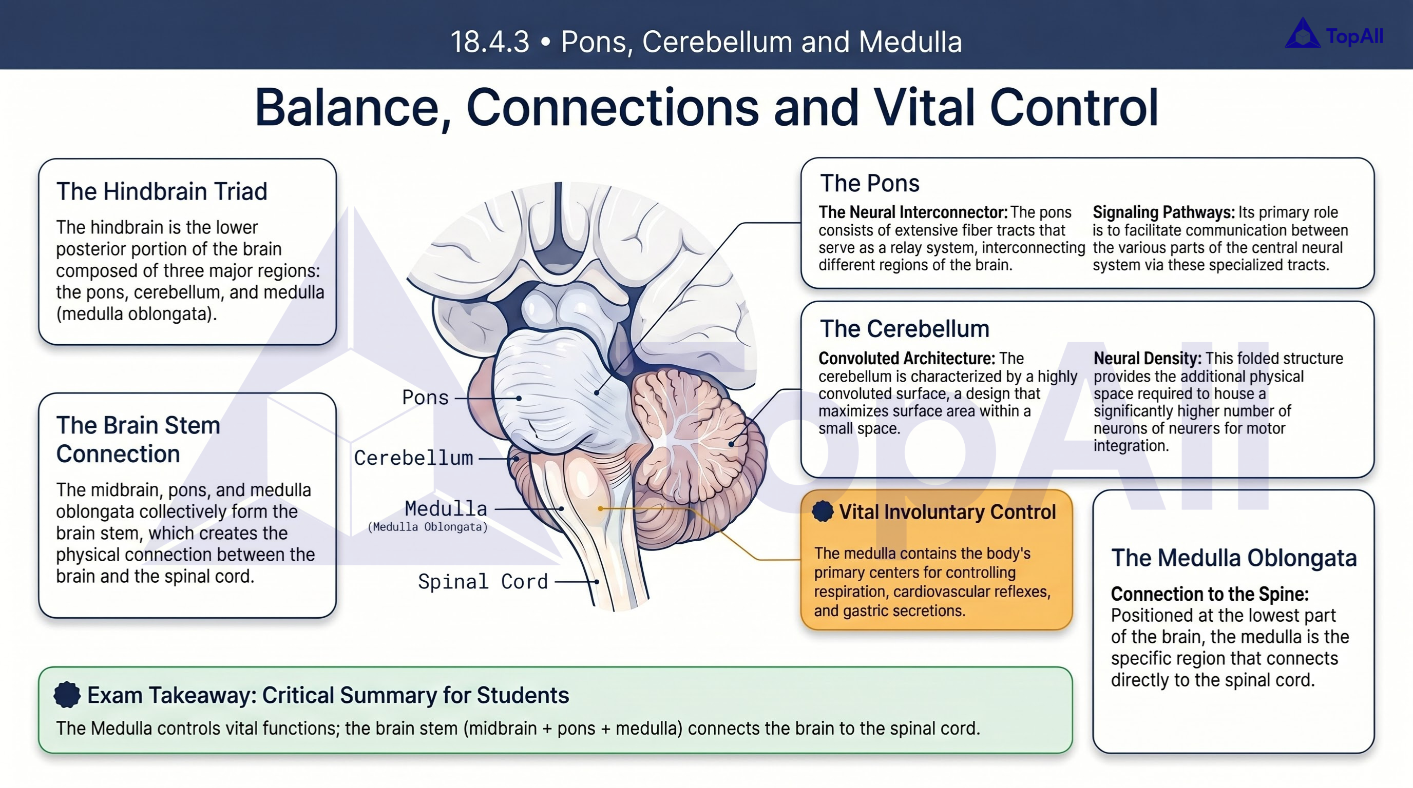
Learn hindbrain parts and functions for NEET UG with TopAll.
NEET UG · Mind map
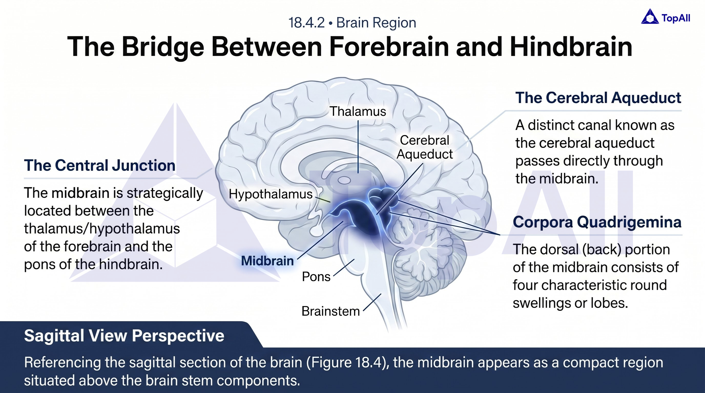
Understand midbrain structure and role in coordination for NEET UG with TopAll.
Follow sensory and motor pathways, distinguish ion-channel flow from pump maintenance and reason through a chemical-synapse release step.
For NEET UG Biology students: trace afferent and efferent signals, distinguish channel-mediated ion flow from pump maintenance and keep axonal conduction separate from transmitter release at a chemical synapse.
Afferent pathways carry information toward the CNS, while efferent pathways carry commands toward peripheral effectors. An action potential propagates along an excitable membrane through changes in ion-channel conductance. At a chemical synapse, a presynaptic signal causes neurotransmitter release and a receptor-dependent response in the target. These are connected steps, but blocking a synaptic release mechanism is not the same event as reversing axonal conduction.
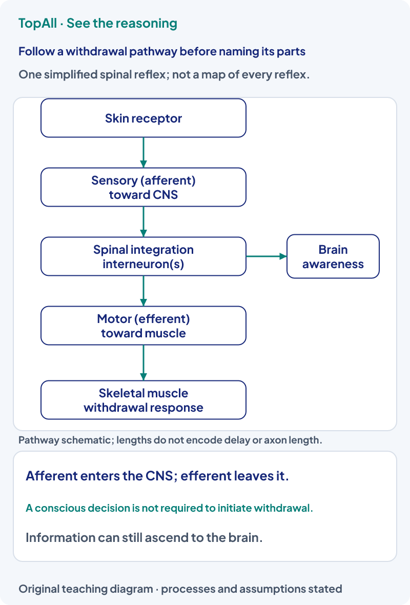
In a diagram, a labelled connection runs from a skin receptor toward spinal processing, and another runs from processing toward a flexor muscle. The first is afferent and the second efferent, regardless of whether their lines point left or right on the page. If only the illustrated afferent connection is completely interrupted, that stimulus cannot reach this reflex pathway through it. An intact efferent connection alone cannot replace the missing sensory input. The statement concerns this specified pathway, not every possible source of muscle activation.
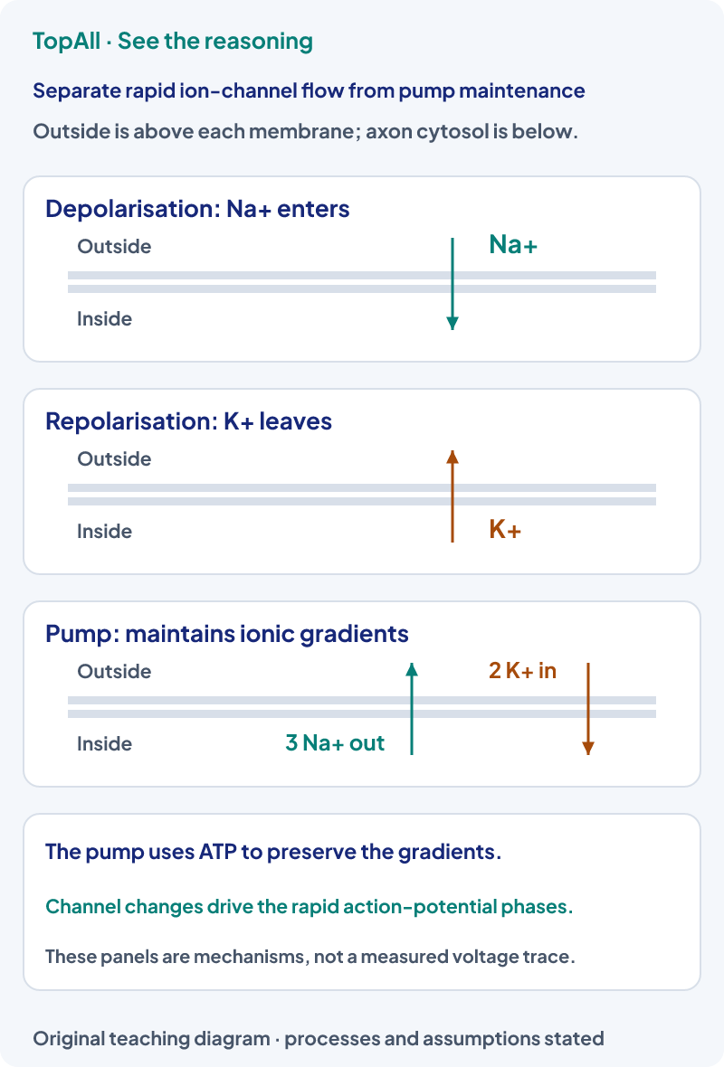
In the resting axon model, Na⁺ concentration is higher outside and K⁺ higher inside. The ATP-dependent sodium-potassium pump moves outward for inward per cycle. Rapid Na⁺ entry through activated channels contributes to depolarisation; Na⁺ channel inactivation and increased K⁺ exit contribute to repolarisation. The pump supports long-term gradients but is not the fast potassium-channel phase itself. A potential difference describes charge separation across the membrane, not a large imbalance throughout all the cell fluid.
Consider a chemical synapse whose axon can still conduct normally, but presynaptic entry at the terminal is prevented. The action potential may reach the terminal, yet the calcium-triggered vesicle-release step is reduced or prevented under that assumption. Less transmitter reaches postsynaptic receptors, so the usual postsynaptic response is impaired. It does not follow that calcium normally carries the entire action potential along the axon. Presynaptic calcium entry and axonal sodium/potassium conductances belong to different mechanistic steps.
At a typical chemical synapse, transmitter is released from the presynaptic side and binds receptors on the postsynaptic side. The effect depends on transmitter, receptor and ion conductances; some responses are excitatory and others inhibitory. The presence of a neurotransmitter does not guarantee a new postsynaptic action potential. Electrical synapses use direct ionic current through intercellular connections and are a different mechanism; do not apply the vesicle-release account to every synapse type.
For a supplied model of four complete Na⁺/K⁺ pump cycles, 4×3=12 Na⁺ move outward and 4×2=8 K⁺ move inward. The net transported charge is four positive elementary charges outward across those cycles. This stoichiometric count does not calculate a whole neuron’s voltage change, action-potential amplitude or total ion traffic. Those depend on membrane properties and channel flows as well as the number and timing of pump cycles.
The CNS comprises brain and spinal cord. The cerebrum participates in sensory, motor and higher processing; the cerebellum helps coordinate movement and balance; medullary centres regulate functions including breathing and cardiovascular reflexes. The hypothalamus links homeostatic and endocrine control. These are functional associations, not a claim that any complex behaviour depends on one isolated structure. In pathway questions, identify the level and connection actually specified.
Label “toward CNS” and “away from CNS” before naming afferent or efferent. Separate axonal propagation, terminal release and postsynaptic response. When ions are shown, name both the side and the mechanism. Interpret an action-potential question for the stated single axon rather than silently replacing it with a recording from a whole mixed nerve.
For the same axon under comparable membrane conditions, a stronger sustained stimulus is applied after threshold has already been exceeded. Must every individual action potential become larger?
No. Individual action potentials are approximately all-or-none under those comparable conditions. Stimulus strength can be represented by firing frequency and, in a larger system, recruitment of additional fibres rather than a bigger action potential in the same axon. A compound recording from multiple fibres is a different question. Refractory periods and adaptation also limit any simple frequency prediction.
A question asks which movement chiefly contributes to the rapid repolarising phase of a typical axon action potential. Choose between K⁺ efflux through channels and the Na⁺/K⁺ pump cycle, and justify the distinction.
K⁺ efflux through the relevant channels contributes to rapid repolarisation, together with reduced Na⁺ conductance as its channels inactivate. The pump uses ATP to maintain gradients over time and has a three-out/two-in stoichiometry. Its maintenance role does not make it the channel-mediated repolarisation event.
Trace sensory input and motor output, then write which membrane mechanism or synapse each step uses. Use Locomotion and Movement to follow the motor signal into skeletal muscle, or Structural Organisation in Animals to distinguish a neuron from the tissues in an organ.
Practice opens the broad Neural Control and Coordination chapter pool, not a guaranteed reflex-only set.
Technical review: Codex (automated), 24 October 2026. Definitions and conventions checked against the linked primary textbooks. Numerical examples, self-checks and diagrams are original; calculations and diagram coordinates are checked separately. Human subject review is not claimed. These focused lessons support the existing NEET Biology chapter catalogue and do not claim complete current exam or syllabus coverage.
After a neural-pathway error, check direction relative to the CNS, membrane compartment and synaptic step independently before returning to chapter practice.